 |
Toll Free 877-238-1437
|
Breast Brachytherapy
( High Dose Rate Brachytherapy for Breast Cancer )
Back to Top1. Introduction
| Over the past two decades, breast conserving treatment, consisting of lumpectomy and external beam radiotherapy, has become a standard treatment option for patients with early stage cancer. It has been demonstrated to offer similar local control and survival results compared with mastectomy, while providing acceptable cosmetic outcome, and less emotional trauma. However, the primary disadvantages are 6 to 7 weeks of external beam treatment and potential side effects to the adjacent organs such as the underlying lungs and scatter radiation dose to the opposite breast. |  |
Back to TopTable of Content
- Introduction
- Advantage of HDR Brachytherapy
- Surgery and HDR Procedure
- Computer Dosimetry
- Cosmetic Result
Back to Top2. The Advantages of HDR Brachytherapy
- The overall treatment time is reduced to 5 to 7 days compared to a 6 to 7 week course of external beam radiotherapy.
- Breast brachytherapy delivers a precisely targeted dose to the tissues most at risk for recurrence, increasing the likelihood of tumor control.
- Reduces radiation dose to the lungs and opposite breast
- Reduces radiation dose to the breast outside the brachytherapy treatment volume, avoiding potential long term adverse cosmetic side effects associated with whole breast external beam therapy, such as fibrosis and skin reactions.
- Breast brachytherapy causes no delays in other treatments such as chemotherapy.
- Conserve your breast and yield excellent cosmetic result .
Back to Top3. Surgery and HDR Procedure
The brachytherapy implant can be done at the time of lumpectomy or later, after the lumpectomy incision has healed. The patient is placed under general anesthesia. The brachytherapy physician places rows of thin hollow tubes, the treatment catheters, to encompass the tumor bed plus generous margins. These catheters will later hold the radioactive source. The catheters are left in place with the catheter ends protruding through the skin for the duration of the treatment course, which is about five days. On the first day of treatment, radiographic or CT images of the implant are obtained for treatment planning purposes. After the radiation dose plan is reviewed and approved by the physician, the treatments can begin. |
The protruding ends of the plastic catheters are connected via longer, transfer tubes to the treatment unit, called the afterloader, that contains the tiny radioactive source. The tiny (1mmx3mm) source is welded to the tip of a flexible stainless steel cable that travels in and out of the treatment catheters. The source steps through each of the catheters, stopping every 5mm to deliver the required radiation dose. After the treatment is delivered, the source retracts into the afterloader, the treatment catheters are disconnected from the transfer tubes, the nurses cover the implant with dressings and the patient goes home. The time the source spends in the catheters is about 10 minutes. |
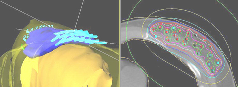
Back to Top4. Computer Dosimetry
| Fig 2: 3D computer simulation showing radiation dose cloud. | ||
 |
||
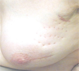
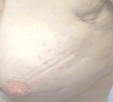
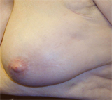
Back to Top5. Cosmetic Result
 |
 |
 |
||||
| Fig 3: 2 months after treatment. | Fig 4: 10 months after treatment. | Fig 5: 3 years after treatment, achieve excellent cosmetic result. | ||||
Home
Our Treatment Programs
- Prostate Cancer

 Monotherapy Survival Rate
- Vaginal Cancer
- Cervical Cancer
- Endometrial Cancer
- Vulvar cancer
- Breast Cancer
- Head and Neck Cancer
- Lung Cancer
- Esophageal and Bile Duct Cancer
- Soft Tissue Sarcoma Cancer
Our Treatments Statistics
Frequently Ask Questions
General Information
Our Publications
Cancer Resources & Links
Back to TopGeneral Frequently Asked Questions
1. How does radiation kill cancer?
Cancer is made of abnormal cells that tend to grow without control. Cancer DNA is more sensitive to radiation than are normal cells, so radiation kills cancer directly or when the cells attempt to multiply while normal tissue in the region is able to repair and recover.
2. What is Brachytherapy?
The prefix "brachy" is the Greek word for "short" distance. Brachytherapy is a form of internal radiation treatment where radioactive sources are placed on or into cancer tissues. There are two kinds of brachytherapy. The radiation sources may be inserted either permanently or temporarily. The two most common forms of treatment are low dose rate (LDR) permanent seeds for prostate cancer and high dose rate (HDR) temporary brachytherapy, that can be used for prostate, gynecologic, breast, head and neck, lung, esophageal, bile duct, anorectal, sarcoma, and other cancers.
3. What is high dose rate (HDR) Brachytherapy?
High dose rate (HDR) is a technically advanced form of brachytherapy. A high intensity radiation source is delivered with millimeter precision under computer guidance directly into the tumor killing it from the inside out while avoiding injury to surrounding normal healthy tissue. For a more in depth explanation please go to understanding HDR Brachytherapy page.
4. What are the advantages of HDR Brachytherapy?
- Preservation of organ structure and function
- Improved accuracy and precision of radiation dose delivery
- Knowledge of radiation dose distribution before treatment is given
- Ability to shape the radiation dose to fit the tumor
- Fewer side effects
- No radiation source (seeds) migration into other organs
- No radiation exposure to other persons
- The treatment course is days rather than weeks to months (as required for permanent seeds or external beam)
- Excellent coverage of possible microscopic extension of cancer
- Minimizes areas of radiation overdose (hot spots) or underdose (cold spots)
- Organ motion (target movement) is not a problem for HDR as it is with external beam
- Effective treatment for cancer recurrence (termed "salvage" therapy)
5. How successful is HDR Brachytherapy?
HDR Brachytherapy is proven to be effective for the treatment of local disease in many forms of cancer including prostate, gynecological, breast, head and neck, esophagus, lung, anorectal, bile duct, sarcoma, and other primary cancer or localized metastasis as reported in the medical literature. CET's publication on prostate cancer, for example has demonstrated 90% 10-year tumor control. Success rates for other tumors vary according to the type and stage of cancer being treated.
6. How many treatments has CET administered?
As of 8/31/2005, CET has performed 8,023 HDR implants and delivered 16,464 HDR treatments. Please see our treatment statistics for further details.
7. Why is HDR less well known than other forms of cancer treatment?
HDR Brachytherapy is a relatively new form of advance radiation technology. Fewer physicians have been trained to perform HDR procedures compared to seed implants or external beam radiation. Few centers, other than CET have been dedicated to the development of HDR brachytherapy to its full potential. Dr. Demanes has devoted his career to the advancement of brachytherapy and has pioneered the use of HDR and established CET as a center of excellence with specially trained and experienced staff and physicians.
8. Why should I select CET?
- Most experienced HDR center in the country
- First center specializing solely in HDR brachytherapy
- Recognized as HDR experts by colleagues in radiation oncology
- Acknowledged safety record
- Highly trained and experienced physicians and staff
- Long term results published in peer reviewed medical literature
- Quality patient care and follow up
Back to TopAbout Us
-
California Endocurietherapy Cancer Center (CET) is the first brachytherapy only center in the United States.
-
Founded by D Jeffrey Demanes M.D. in 1981.
-
Dedicated solely to High Dose Rate brachytherapy (HDR) since 1991.
-
Most experienced HDR brachytherapy center.
-
A training destination for physicians and residents.
-
HDR treatment protocol development
-
Innovation in high dose rate brachytherapy and equipment design
-
Dedicated to long-term follow-up, outcome studies, and publications in medical journals.
Membership and affiliations
 |
American Society for Therapeutic Radiology And Oncology Chair - Health Policy and Economics Practice Management Subcommittee, Chair - Regulatory Subcommittee, Member - Health Policy and Economic Committee, Member - Health Policy and Economics Code Development and Valuation Subcommittee, Member - Code Utilization and Application Subcommittee. |
 |
|
 |
American College of Radiation Oncology President - 2005 to 2006 |
Back to Top
CET Cancer Center
California Endocurietherapy Medical Corp.3012 Summit St. Suite 2675, Oakland, CA 94609
Tel: 877-238-1437 or 510-986-0690 Fax: 510-986-0159
Hours: Mon-Fri, 8:30AM to 5:00PM Pacific Time
Copyright California Endocurietherapy Medical Corp. All Rights Reserved. www.cetmc.com